


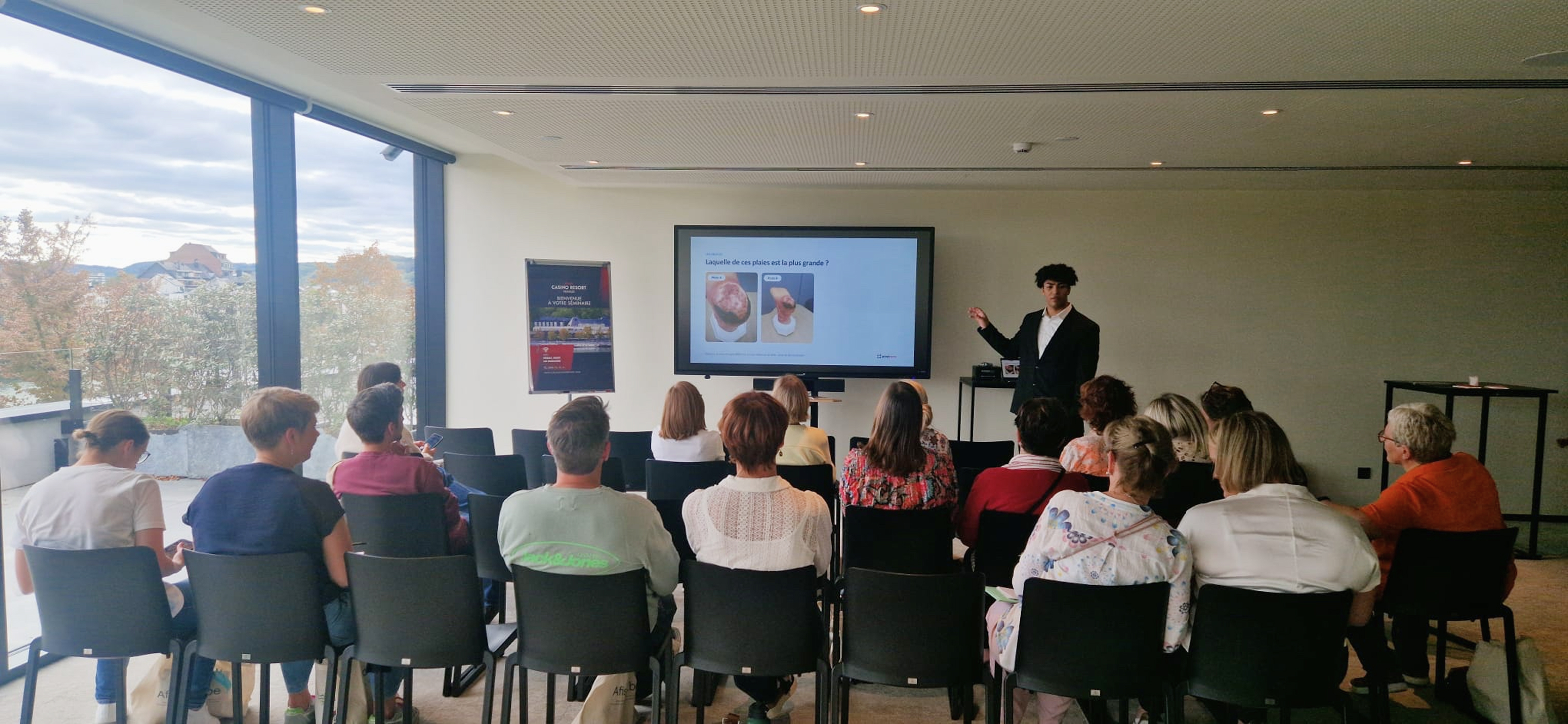
Every clinician photographs wounds. Very few of those photos become reliable data. That was the starting point of the workshop Pixacare presented on 15 October 2026 at the AFISCEP refresher congress in Belgium: ‘The uses of AI and photography apps: challenges, uses, limitations, outlook’.
This article covers the key points: what AI can really do with a wound photo, what it cannot see, and five simple questions to assess any wound measurement or analysis tool before adopting it in your service.
Wound photography: a photo is not yet a measurement
Photography is already part of care: it is used to monitor how a wound progresses, share information between home, community and hospital care, get a remote opinion and record what was done. But a photo taken without a method can be misleading.
During the workshop, the audience compared two photos and had to say which wound was larger. It was the same wound, photographed on the same day, a few minutes apart. Only the distance, the lighting and the angle had changed. Without a size reference, the eye cannot tell.

Why wound measurement matters from week 4
A landmark study on diabetic foot ulcers (Sheehan et al., Diabetes Care, 2003) measured wounds at baseline and again at 4 weeks:
- when the surface area had decreased by more than 53%, 58% of wounds had healed at 12 weeks;
- otherwise, only 9% had.
At 4 weeks, you can already tell which wounds are at risk of poor healing, provided they are measured correctly. The length × width ruler method overestimates the area of irregular wounds, and two clinicians rarely get the same figure. This is where digital tools promise to help: turning an image into comparable data.
AI and wounds: four families of tools behind one word
The term AI covers very different tools. Before judging a tool, the first question is simple: what exactly does the tool do?
Measurement
What the tool does: outline, surface area, length and width from a photo with a calibration marker.
Limitation to keep in mind: no depth on a 2D photo; depends on how the photo is taken.
Examples: WoundTrack II (Pixacare), imitoWound, Minuteful for Wound (Healthy.io), Swift Skin and Wound, eKare inSight; in 3D: ARANZ Silhouette, cureVision.
Tissue analysis
What the tool does: proportion of granulation tissue, slough and necrosis.
Limitation to keep in mind: sensitive to lighting and skin tone; outline to be checked.
Examples: WoundTrack II and several of the tools above, Tissue Analytics (Net Health).
Specialised imaging
What the tool does: dedicated device for bacterial fluorescence or multispectral imaging.
Limitation to keep in mind: specific hardware, targeted use.
Examples: MolecuLight (fluorescence), Kent Imaging (oxygenation), DeepView by Spectral AI (burns).
Generative AI
What the tool does: writing a handover note, summarising a record, answering a question.
Limitation to keep in mind: can make up an answer; no patient data in a consumer tool.
Examples: ChatGPT, Le Chat, Copilot.
Not to be confused: wound monitoring and case-sharing platforms (secure photo records, progress curves, remote specialist advice) are not AI. They are very useful, and AI is often built into them. At Pixacare, the monitoring platform and the AI analysis module are in fact two separate medical devices, in different classes.
The limitations of AI applied to wound photos
An AI is only as good as the photo it is given and the data it learnt from. Three tricky photos shown during the workshop illustrate this well:
- Flash: its glare whitens the wound bed, which an algorithm may read as slough, and hides the true edge;
- Darker skin tones: the contrast between skin and wound is lower, and an algorithm trained mostly on lighter skin may outline it less accurately;
- Cavity wounds: a photo is two-dimensional, so it sees neither depth nor what lies beneath the edges.

Beyond these pitfalls, the limitations fall into five categories:
- Technical: lighting, angle, distance; deep, tunnelling or circumferential wounds.
- Bias: sometimes lower performance on darker skin tones if they are under-represented in the training data.
- Clinical: the image says nothing about pain, odour, warmth or the patient's context.
- Data: personal phone, mixed photo gallery, consumer messaging apps: data protection law (UK GDPR or EU GDPR) applies to every photo.
- Organisational: training time, duplicate data entry if the tool is not connected to the patient record.
The real risk is not that AI gets it wrong. It is that we stop looking because it gave us a number.
CE marking and the AI Act: the European regulatory framework for wound software
In the European Union, software that measures or analyses a wound generally falls under the EU Medical Device Regulation (EU MDR 2017/745). It then carries a CE mark and a risk class. But not all CE marks are equal:
- Class I: the manufacturer self-declares conformity, with no external audit (except for sterile, measuring or reusable devices);
- Class IIa and above: an independent notified body audits the device, and its four-digit number appears next to the CE logo.
So the first reflex is to check whether there is a number next to the CE logo.
A second layer is coming: the EU AI Act. For medical devices that include AI, the requirements for high-risk AI systems, including transparency and human oversight, will apply from 2 August 2028, following the postponement adopted under the ‘Digital Omnibus’ regulation.
5 questions to ask before adopting an AI tool for wound care
This checklist was presented during the workshop. It applies to any tool, ours included.
- Is it CE marked as a medical device, and in which class? Class I: self-declared by the manufacturer. Class IIa and above: audited by a notified body, whose number follows the CE logo.
- Where are the photos hosted, and who can access them? Secure hosting suited to health data, role-based access, nothing in the phone's photo gallery or on a consumer messaging app.
- Which patients and which wounds was it validated on? Wound types and sizes covered, skin types represented, published studies.
- Does it integrate with the patient record, without duplicate data entry? Connection to the EPR, report export, secure sharing between home, community and hospital care.
- Who keeps the decision, and how does the tool remind users of it? The tool measures and analyses. The clinician checks, corrects and decides.

Pixacare and WoundTrack II answer their own questions
Pixacare was founded in 2019 in Strasbourg, France, and co-founded by Prof. Frédéric Bodin, a surgeon at Strasbourg University Hospital. The platform is used by more than 70 healthcare facilities in 6 countries. Here is how it answers the checklist.
- CE marking and class: WoundTrack II, the AI wound analysis module: Class IIa, CE 2460 (notified body DNV) since March 2026. Pixacare Application: Class I. Quality management system certified to ISO 13485:2016.
- Hosting and access: data hosted in France, role-based access, SSO available, photos kept out of the personal gallery.
- Validation: chronic wounds in adults, studies detailed below.
- Integration with the patient record: API, HL7, FHIR R4, PDF reports to the EPR.
- Who decides: the clinician. The AI suggests an outline and a tissue breakdown, which the clinician checks and can correct.
And what the tool does not do: it does not measure depth, and it analyses one wound per photo. Above all, it does not replace the clinician's clinical judgement.
What the clinical studies show
- Wessels, Berkane, Bodin, ETRS 2026 (publication forthcoming): 100 photos, AI corrected by the clinician, vs 3 clinicians using planimetry. Key result: median error 4.7%; ICC 0.993.
- Pixaire1, Maxant et al., 2026 (medRxiv): 36 patients, semi-automated measurement vs planimetry. Key result: ICC 0.96; limitations: wounds on several planes, blurred edges.
An intraclass correlation coefficient (ICC) close to 1 means near-perfect agreement with the reference measurement. In the study presented at ETRS 2026, this level is reached by AI that the clinician has reviewed and corrected: the clinician stays in the loop.
3D measurement, trajectory tracking: what's next
Several trends are emerging in the sector, beyond the uses currently covered by certified devices. These are research and development directions, not features available today:
- 3D measurement directly with smartphone depth sensors, without a dedicated device;
- measurement without a calibration sticker, so patients can photograph their own wound at home;
- trajectory tracking, to flag earlier a wound whose progress departs from the expected curve, with the decision remaining with the clinician;
- linking wound analysis to the organisation's dressing protocol, with the clinician keeping the final choice.
Each of these advances will need clinicians: to take good-quality photos, to validate tools in real-world settings, and to point out what the machine cannot see.
Frequently asked questions about AI and wound photography
Can AI measure a wound from a simple photo?
Yes, provided the photo includes a calibration marker. The AI then detects the outline and calculates the surface area, length and width. It does not measure depth on a two-dimensional photo.
Does a wound photo app need to be CE marked?
In the EU, software that measures or analyses a wound generally falls under EU MDR 2017/745 and must carry a CE mark as a medical device. From Class IIa upwards, an independent notified body has audited it, and its number appears next to the CE logo.
Does AI work equally well on all skin tones?
Not always. On darker skin tones, the contrast between skin and wound is lower, and an algorithm trained mostly on lighter skin may outline the wound less accurately. That is why you should ask which skin types the tool was validated on.
Does AI replace the clinician's assessment?
No. AI measures and analyses; the clinician observes, interprets and decides. With WoundTrack II, the AI suggests an outline and a tissue breakdown that the clinician checks and can correct.
Three key takeaways
- One standardised photo is worth more than ten random ones.
- Before adopting a tool, ask the 5 questions.
- AI measures and analyses; the clinician observes, interprets and decides.
Would you like to try WoundTrack II in your service? Contact the Pixacare team.
Références
- Sheehan P, Jones P, Caselli A, Giurini JM, Veves A. Percent change in wound area of diabetic foot ulcers over a 4-week period is a robust predictor of complete healing in a 12-week prospective trial. Diabetes Care. 2003;26(6):1879-1882. doi:10.2337/diacare.26.6.1879
- Wessels, Berkane, Bodin. AI-based chronic wound area measurement corrected by the clinician vs planimetry. Presentation, ETRS 2026 congress (publication forthcoming).
- Maxant G, Mori C, Maxant T, Bertaux AC. Pixaire1: Evaluation of automated chronic wound surface measurement systems. medRxiv, 31 March 2026 (preprint). doi:10.64898/2026.03.30.26344793
- Regulation (EU) 2017/745 of the European Parliament and of the Council of 5 April 2017 on medical devices (EU MDR). EUR-Lex
- Regulation (EU) 2024/1689 of the European Parliament and of the Council of 13 June 2024 laying down harmonised rules on artificial intelligence (AI Act). EUR-Lex
- Regulation (EU) 2026/1744 of 8 July 2026 (‘Digital Omnibus’ on AI), published in the Official Journal of the EU on 24 July 2026, postponing to 2 August 2028 the application of the requirements for high-risk AI systems embedded in medical devices. EUR-Lex
- Regulation (EU) 2016/679 of 27 April 2016 on the protection of personal data (GDPR). EUR-Lex


