
Standardize healing documentation

A unified digital protocol to structure photos, questionnaires, measurements and reports, and ensure reliable documentation in accordance with best practices.

Injury documentation is at the heart of clinical decision making

A structured framework for evaluating the evolution of lesions, coordinating care and sharing reliable information throughout the patient journey.

A 100% medical gallery

Structured documentation makes it possible to objectively observe the evolution of the lesions and to ensure continuous clinical follow-up.

Save time during consultations

Organize your photos and videos in seconds.


Clinical follow-up over time

Clear, comparable and structured information facilitates the adaptation of care and therapeutic decision-making.

Essential features to automate medical photo management

Pixacare harmonizes photos, questionnaires, measurements, remote monitoring and reports in a single repeatable flow that is accessible to the entire team.

1

Standardizing the taking of medical photos

Produce reliable, comparable and usable images over time.

Photographic history structured by patient
Chronological comparison of lesions
Centralization of photos taken by various caregivers
Learn more about the medical photo library
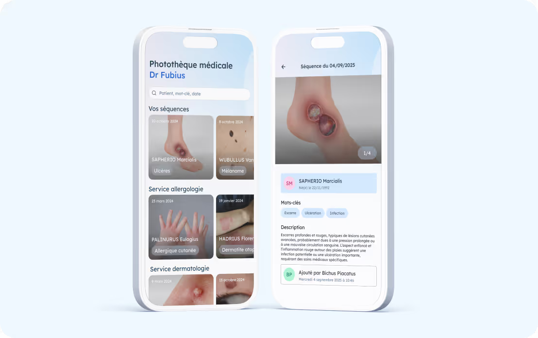
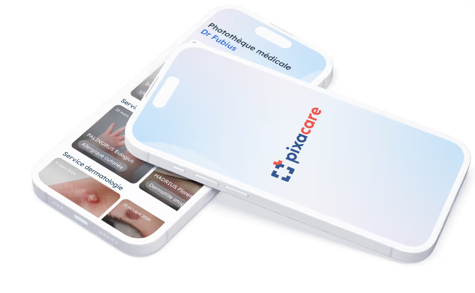
Pixacare application showing a medical photo library with patient sequences for allergology and dermatology, and a detailed view of a red and ulcerated skin lesion on one foot.
2

Structuring clinical evaluations

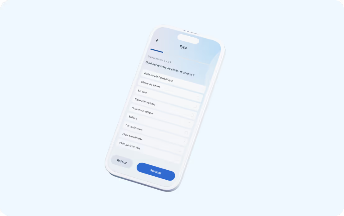
Collect consistent data, adapted to each type of lesion.
Standardized questionnaires in accordance with the recommendations of the HAS
Integration of scores, classifications and standards from reference learned societies
Possibility to create personalized questionnaires, according to service practices
Pixacare application displaying a questionnaire on the type of chronic wound with various options to select.

Examples of clinical questionnaires available, based on the recommendations of the HAS and reference scientific societies.

Complex chronic wounds

TIME, MEASURE, TILI, Healing Triangle, BWAT
Close-up of a post-operative surgical wound with visible stitches on the skin.

Postoperative wound

IDE, PRADO, HAD and SSIA protocols checklist
Nurse in a blue uniform holding the hand of an elderly woman sitting on a sofa, in a warm setting.

HAD

PRADO
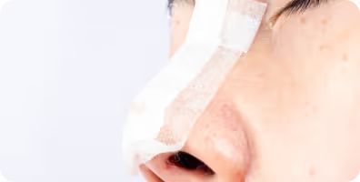
Close-up of a face with a medical bandage on the nose, indicating plastic or reconstructive surgery.

Plastic and reconstructive surgery

FACE-Q, BREAST-Q, BODY-Q, SCAR-Q, POSA
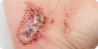
Scar with blue stitches and a transparent drain on the skin.

Hand surgery

DASH, PRWHE, TAM
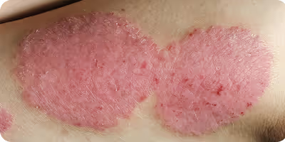
Two red, scaly patches of atopic dermatitis on the skin.

Atopic dermatitis and eczema

SCORAD, EASI, POEM, NRS-Pruritus
3

Measure wounds accurately and reproducibly usingartificial intelligence

Depending on the context, two methods are possible:
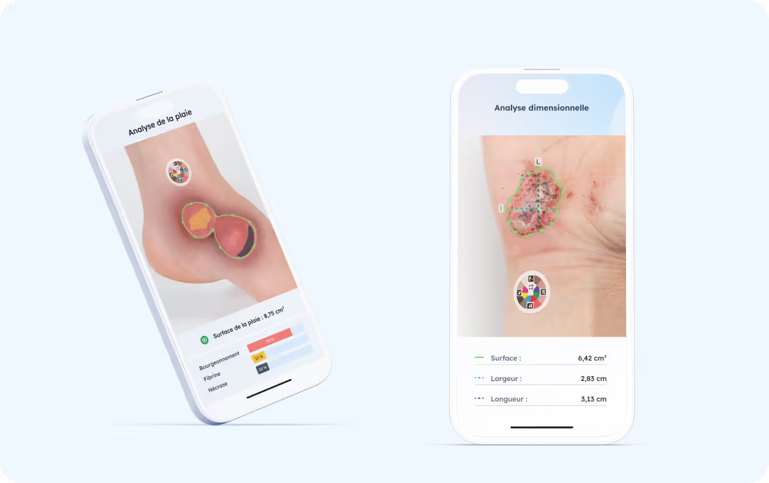
Automatic measurement assisted by AI: surface + tissue analysis.
Manual planimetry: area, lengths, segments.
Automatic correction of distances and colors thanks to the patented sticker.
Learn more about automatic measurements
Two smartphones displaying the Pixacare wound analysis application with area, width and length measurements on images of injured skin.
4

Integrating telemonitoring into the care pathway

Document healing in the home without breaking clinical continuity.
Reception of photos, questionnaire responses and measurements taken at the patient's home.
Automatic integration into the patient file.
Easy to use for patients or IDE in the city.
Learn more about remote tracking
Pixacare application displaying the Pixacare logo above a blue cloud icon, symbolizing cloud sync.
5

Generate a complete healing report

Valorize the data collected in a structured, graphic and easily shareable format.
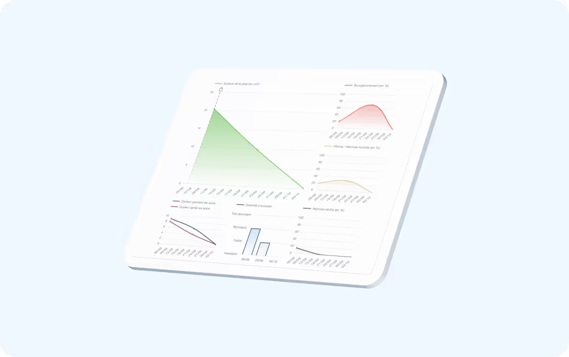
Reports containing photos, measurements and graphical visualizations.
Assessments facilitate therapeutic adaptation, longitudinal follow-up, as well as reimbursement claims and quality audits.
Automatic PDF export to the DPI
See a report
Mockup of the Pixacare application displaying a healing report with several colored graphs on skin surface evolution, buds, pain, and other measures.

Frequently asked questions about the clinical documentation of lesions

Puis-je tester Pixacare avant de l'adopter ?

Les rapports de cicatrisation peuvent-ils être intégrés au DPI ?

Comment sont générés les bilans de cicatrisation ?

Peut-on personnaliser les questionnaires cliniques ?

Deploy the medical image library